


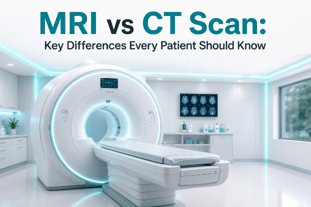
You've just been asked to get an MRI or a CT scan — but do you know the difference? Both are powerful diagnostic imaging tools, yet they work in completely different ways, reveal different types of information, and are suited to different medical situations. This guide demystifies both scans so you can walk into your next appointment fully informed.
What is an MRI Scan?
Magnetic Resonance Imaging (MRI) uses powerful magnetic fields and radio waves to generate detailed images of the body's soft tissues, organs, and internal structures. There is no ionizing radiation involved, making it a preferred option for repeated use and for imaging sensitive areas like the brain and spinal cord.
During an MRI, the patient lies inside a large cylindrical machine. A strong magnetic field aligns hydrogen atoms in the body, while radio wave pulses generate signals that are captured and processed into detailed cross-sectional images. The process is entirely non-invasive and painless — though it does produce loud knocking sounds and can feel claustrophobic for some patients.
Key Technology: MRI uses magnetic fields + radio waves. No radiation. Exceptional detail in soft tissues — brain, spinal cord, ligaments, cartilage, and internal organs.
What is a CT Scan?
Computed Tomography (CT), also called a CAT scan, uses X-rays taken from multiple angles to create cross-sectional "slices" of the body. A computer then assembles these slices into a detailed 3D image. CT scans are significantly faster than MRIs and are particularly effective for imaging bones, detecting internal bleeding, and evaluating trauma or emergency conditions.
The procedure involves the patient lying on a motorized table that moves through a ring-shaped scanning machine. It takes only a few minutes — sometimes under 30 seconds for the actual scan. A contrast dye may be injected to make blood vessels, tumors, or organs more visible.
Key Technology: CT uses X-ray beams from multiple angles. Fast scanning. Excellent for bones, lung tissue, blood vessels, and trauma assessment. Does involve low-dose radiation.
MRI vs CT Scan: Side-by-Side Comparison
Here's a quick comparison of the key differences between MRI and CT scans across important parameters:
| Feature | MRI | CT Scan |
|---|---|---|
| Technology | Magnetic fields + radio waves | X-ray beams (multiple angles) |
| Radiation | None | Low-dose ionizing radiation |
| Scan Duration | 20–90 minutes | 5–10 minutes |
| Best For | Soft tissues, brain, spine, joints | Bones, lungs, trauma, internal bleeding |
| Image Detail | Superior soft tissue contrast | Superior bone and dense tissue detail |
| Noise Level | Very loud (knocking sounds) | Quiet |
| Claustrophobia Risk | Higher (enclosed bore) | Lower (wider, open ring) |
| Use in Pregnancy | Generally considered safer | Avoided unless critical |
| Metal Implants | Contraindicated (pacemakers, certain implants) | Generally safe (with precautions) |
| Cost (India approx.) | ₹4,000 – ₹25,000+ | ₹1,500 – ₹10,000+ |
| Emergency Use | Limited (slower, not for unstable patients) | Preferred (fast, widely available) |
| AI-Assisted Reading | Available via RadpicsAI | Available via RadpicsAI |
When is an MRI Recommended?
Doctors typically order an MRI when they need a highly detailed look at soft tissues — structures that don't show up clearly on X-rays or CT scans. MRI is the gold standard for neurological and musculoskeletal conditions.
MRI is typically ordered for:
- Brain tumors and lesions
- Multiple sclerosis (MS)
- Spinal cord injuries
- Stroke evaluation
- Knee, shoulder, and joint damage
- Torn ligaments and tendons
- Liver and kidney diseases
- Pelvic conditions (uterus, ovaries, prostate)
- Breast cancer evaluation
- Vascular abnormalities (MRA)
When is a CT Scan Recommended?
CT scans shine in emergency and trauma settings where speed is critical. They are also the preferred tool for imaging structures like bone, lungs, and blood vessels, or when metal implants preclude MRI use.
CT Scan is typically ordered for:
- Head injuries and skull fractures
- Internal bleeding detection
- Chest and lung conditions
- Pulmonary embolism
- Abdominal emergencies
- Kidney stones
- Bone fractures and tumors
- Cancer staging and monitoring
- Appendicitis diagnosis
- Cardiovascular assessment (coronary CT)
Radiation: Should You Be Concerned?
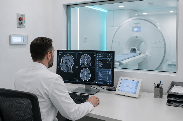
One of the most common concerns patients raise is radiation exposure. Here's what you need to know: MRI uses no ionizing radiation at all. It is entirely magnetic — safe for children, pregnant women (in most cases), and patients requiring multiple follow-up scans.
CT scans do emit ionizing radiation, though at much lower levels than older machines thanks to modern dose-reduction technology. A typical chest CT exposes a patient to roughly 7 millisieverts (mSv) — comparable to about 2 years of natural background radiation. For most adults, the diagnostic benefit far outweighs this minimal risk. However, repeated CT scans over time should always be discussed with your physician.
Important: Children and pregnant patients should avoid CT scans unless medically essential. Always inform your doctor if you are pregnant or may be pregnant before any imaging procedure.
Key Factors That Influence the Choice
- Type of Tissue: Soft tissue such as the brain, muscles, and ligaments points toward MRI. Bone, calcification, and dense structures are better evaluated with CT.
- Urgency: Emergency or trauma situations call for CT due to speed. Elective or detailed soft-tissue evaluation is better served by MRI.
- Metal Implants: Patients with a pacemaker, cochlear implant, or certain metal devices may find CT safer, as MRI can be contraindicated in these cases.
- Pregnancy: MRI is preferred during pregnancy since it involves no radiation. CT is used only when medically essential.
- Claustrophobia: CT machines are wider and less enclosing than traditional MRI scanners. Open MRI options are also available for anxious patients.
- Cost and Availability: CT scans are generally faster, more affordable, and more widely available — particularly in smaller diagnostic facilities.
Can You Have Both?
Yes — and in complex cases, doctors often order both. For example, a cancer patient may receive a CT scan for initial staging (fast, full-body overview) followed by an MRI for a detailed assessment of a specific tumor's involvement in surrounding soft tissue. The two scans provide complementary information rather than competing findings.
Advanced AI-powered platforms like RadpicsAI's MedVIEWER™ allow radiologists to analyze both MRI and CT images in a unified viewer — with AI-assisted annotations, measurements, and structured reporting — delivering faster, more accurate diagnostic insights to treating physicians.
What Should You Ask Your Doctor?
Walking into your appointment informed makes a real difference. Here are the right questions to raise:
- Why is this specific scan being recommended for my condition?
- Are there alternatives, and what are the trade-offs?
- Will contrast dye be used? Am I allergic to iodine or gadolinium?
- I have a metal implant — is MRI still safe for me?
- How long will the scan take, and what should I expect during the procedure?
- Will my results be reviewed by an AI-assisted system, and how quickly will I receive them?
How RadpicsAI Enhances Both MRI and CT Diagnostics
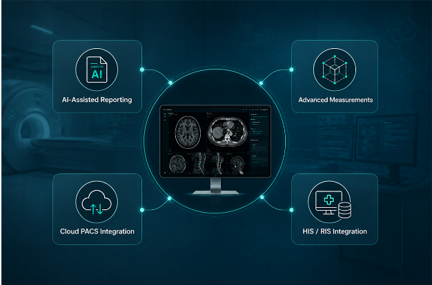
At RadpicsAI, we believe that better imaging technology should translate directly into better patient outcomes. Our AI-powered platform — MedVIEWER™ — supports both MRI and CT scan workflows with:
- AI-Assisted Reporting — Automated anomaly detection and structured radiology reports that reduce turnaround time by up to 40%.
- Advanced Measurements — MPR, 3D reconstruction, and precision annotation tools for both CT and MRI images.
- Cloud PACS Integration — Seamless access to patient imaging archives from anywhere — hospital, diagnostic centre, or remote location.
- HIS / RIS Integration — Connects your imaging workflow with hospital and radiology information systems for a unified, complete patient record.
Final Thoughts
Both MRI and CT scans are remarkable tools — each with a specific set of strengths. MRI offers unparalleled detail in soft tissue imaging without any radiation risk. CT delivers speed, clarity in trauma situations, and excellent bone imaging with minimal radiation dose. Your doctor's recommendation will always be guided by your specific clinical need.
What matters most is that your scans are read accurately and quickly. That's where AI-assisted radiology platforms like RadpicsAI are transforming the diagnostic landscape — ensuring every image is analyzed with precision, every report generated faster, and every patient receives the care they deserve.
Imaging decisions should always be made by qualified healthcare professionals based on your individual clinical condition.
Frequently Asked Questions
What is the main difference between an MRI and a CT scan?
The core difference lies in the technology used. MRI uses magnetic fields and radio waves to produce detailed images of soft tissues without any radiation. A CT scan uses X-rays from multiple angles to create cross-sectional images and is faster, making it ideal for emergencies and bone imaging. Your doctor will recommend one based on what part of the body needs examination and how quickly results are needed.
Which scan is safer — MRI or CT?
MRI is generally considered safer for long-term or repeated use because it involves zero ionizing radiation. CT scans involve low-dose radiation, which is acceptable for most adults in a single or infrequent setting. For children and pregnant women, MRI is strongly preferred unless a CT is medically unavoidable.
Is an MRI more accurate than a CT scan?
Neither is universally more accurate — accuracy depends entirely on what is being examined. MRI provides superior detail for soft tissues, the brain, spinal cord, joints, and organs. CT is more accurate for bones, lung tissue, internal bleeding, and trauma. In many complex cases, both scans are used together for a complete picture.
Why is an MRI so much louder than a CT scan?
The loud knocking and banging sounds during an MRI are caused by the rapid switching of electrical currents in the gradient coils inside the machine. These coils are responsible for creating the precise magnetic field variations needed to produce images. CT scans, which use rotating X-ray beams, are significantly quieter in comparison. Patients undergoing MRI are typically given earplugs or headphones.
Can I have an MRI if I have a metal implant?
It depends on the type of implant. Many modern implants — such as titanium bone plates and some joint replacements — are MRI-compatible. However, pacemakers, cochlear implants, certain aneurysm clips, and older metallic devices may be unsafe in a magnetic field. Always inform your radiologist and doctor about any implant, device, or metallic object in your body before scheduling an MRI.
How long does each scan take?
A CT scan is typically completed in 5 to 10 minutes, with the actual scanning often taking under 30 seconds. An MRI takes considerably longer — anywhere from 20 minutes to 90 minutes depending on the body part being scanned and whether contrast is used. Functional or advanced MRI protocols can take even longer.
Do CT scans or MRIs require any special preparation?
For most scans, you may be asked to fast for a few hours beforehand, particularly if contrast dye is being used. You will need to remove all metal objects, jewelry, and hearing aids before an MRI. Inform your doctor of any allergies, kidney conditions, or medications, as these can affect the use of contrast agents. Your imaging center will provide specific preparation instructions before your appointment.
Which scan is more expensive — MRI or CT?
MRI scans are generally more expensive due to the complexity of the technology and longer scan times. In India, CT scans typically range from ₹1,500 to ₹10,000 depending on the body part and facility. MRI scans generally range from ₹4,000 to ₹25,000 or more. Costs vary based on location, the type of scan, whether contrast is used, and whether the facility is private or government-run.
Can pregnant women get an MRI or CT scan?
MRI is considered the safer imaging option during pregnancy, particularly in the second and third trimesters, as it does not involve radiation. CT scans are generally avoided during pregnancy due to radiation exposure to the developing fetus, but may be used when the medical situation is urgent and no alternative exists. Contrast agents are typically avoided in pregnant patients unless absolutely necessary. Always consult your doctor before any scan during pregnancy.
How does RadpicsAI improve the MRI and CT reporting process?
RadpicsAI's MedVIEWER™ platform uses AI-assisted analysis to help radiologists read MRI and CT scans faster and with greater consistency. The system supports automated anomaly detection, structured report generation, MPR and 3D visualization, and seamless integration with hospital PACS, RIS, and HIS systems. This translates to faster turnaround times, reduced manual errors, and ultimately better outcomes for patients across hospitals and diagnostic centres.