


When a teleradiology operation has image loading problems, the first instinct is always to upgrade the internet connection. Get faster broadband. Increase bandwidth. Throw more connectivity at the problem.
Sometimes that helps. Often it does not. Because the real bottleneck is not how much data the connection can carry. It is how much data is being sent in the first place.
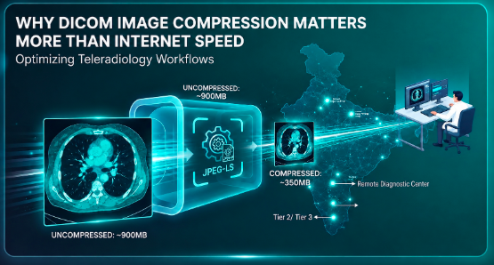
A modern chest CT can contain 600 images. An uncompressed CT study averages 900 MB or more. Sending that over a 12 Mbps connection which is a reasonably good connection in many Indian Tier 2 cities takes over ten minutes. The same study with proper DICOM compression applied can transmit in six minutes or less. That is not a marginal improvement. Across 50 teleradiology reads per day, it is hours.
DICOM image compression is one of the most impactful and least discussed optimisation levers in medical imaging. Here is how it works, why it matters, and how to use it without compromising diagnostic quality.
What is DICOM compression?
DICOM is the international standard for storing and transmitting medical images. Every CT, MRI, X-ray, and ultrasound your hospital produces is a DICOM file a package containing the image data itself plus a header of metadata about the patient, the study, the modality, and dozens of other fields.
In its raw, uncompressed form, a DICOM image file is large. Very large. The image data is stored at full resolution with every pixel value recorded precisely. That is ideal for archiving and diagnostic reading but not ideal for transmission across a network or for efficient storage at scale.
DICOM compression is the process of applying an algorithm to reduce the size of that image file before it is transmitted or stored. The DICOM standard defines which compression formats are acceptable including JPEG, JPEG-LS, and JPEG 2000 so that receiving systems can reliably decompress and display the images.
Lossless vs. lossy: the decision that determines everything
This is the most important distinction in DICOM compression, and it is worth taking a moment to understand it clearly.
Lossless compression
Lossless compression reduces the file size without discarding any image data. When the file is decompressed, it is pixel-for-pixel identical to the original. No information is lost. This is the safe default for diagnostic imaging the radiologist is looking at exactly what the scanner captured.
The common lossless compression formats in DICOM are JPEG-LS and JPEG 2000 lossless. They typically achieve compression ratios of around 2:1 to 3:1, meaning a 900 MB study becomes 300 to 450 MB. That is meaningful bandwidth saving with zero diagnostic compromise.
Lossy compression
Lossy compression achieves much higher compression ratios sometimes 10:1 or more by permanently discarding some image data. The compressed file is smaller, but it cannot be decompressed back to the exact original. Some pixel information is gone.
For primary diagnostic reading, lossy compression is generally avoided. The risk of discarding diagnostically relevant information a subtle finding in a region of the image that was compressed aggressively is not acceptable when a radiologist is making clinical decisions from that image.
However, lossy compression has legitimate use cases. For long-term archiving of studies where the primary diagnostic read has already been completed, some regulatory frameworks permit lossy compression of older studies to reduce storage costs. For patient-facing image sharing where full diagnostic resolution is not needed, lossy compression can make images load faster on mobile devices.
The key principle: never apply lossy compression to images that have not yet been read diagnostically. And always know which compression has been applied to any image you are reading.
Why this matters more than bandwidth
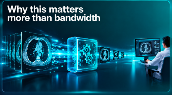
Here is the practical arithmetic that makes compression so impactful.
A radiologist reading 50 CT studies per day, each averaging 900 MB uncompressed, is transferring 45 GB of image data per shift. On a 20 Mbps connection good by most standards that transfer takes roughly five hours of pure data time. Radiologists are not waiting five hours, of course, because studies load progressively. But they are experiencing delays that accumulate throughout the shift.
Apply JPEG-LS lossless compression at a 2.5:1 ratio, and that same 45 GB becomes 18 GB. The same connection now handles the same workload in two hours of data transfer time. The radiologist's studies load noticeably faster. The teleradiology network handles more concurrent sessions without degradation. The cloud storage bill is smaller.
Doubling the internet bandwidth achieves a similar result at ongoing cost, and only until the volume grows again. Compression is a one-time configuration that scales with the volume.
Where compression should happen in the workflow
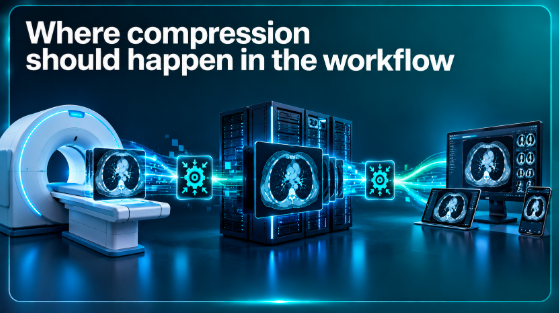
The timing of compression in the imaging chain matters as much as the compression itself.
At the modality (scanner)
Some modalities can apply compression as they generate images. This is the earliest possible point and reduces file size before the image even enters the PACS. However, not all scanners support this, and the compression options available at the modality level are often limited.
At the PACS ingestion point
The most common and controllable compression point is when images are received by the PACS from the modality or from an external source. RadpicsAI's MedPACS applies configurable compression at ingestion — lossless by default for new studies, with separate rules available for archived or imported studies. This happens automatically, without any manual step, as part of the image routing workflow.
At retrieval for viewing
Some PACS platforms apply additional compression when images are retrieved for display serving a lighter version to the viewer for initial loading, then providing the full resolution image on demand. This is particularly useful for teleradiology and patient-facing viewing where the first priority is speed of access, with full resolution available when needed.
The India-specific context
DICOM compression matters everywhere. But it matters especially in India, and for two reasons that are specific to how Indian radiology actually operates.
First, Tier 2 and Tier 3 cities where teleradiology is growing fastest often have real connectivity constraints. A diagnostic centre in a smaller city may have a 10 Mbps or 15 Mbps connection, not a 100 Mbps one. Compression is not optional in that environment; it is what makes cloud PACS workable at all.
Second, the growth of teleradiology networks serving multiple remote centres from a central reading group means that the same images are being transmitted multiple times from the diagnostic centre to the cloud, from the cloud to the reading radiologist, and sometimes back to the referring doctor or the patient. Compression at each stage compounds the benefit.
RadpicsAI's MedPACS was designed with Indian connectivity realities in mind. Compression is applied at ingestion, transmission is optimised for variable bandwidth, and the viewer uses progressive loading starting with a lower resolution preview that loads quickly, then filling in full resolution as the connection allows. Radiologists see something useful immediately, rather than waiting for a full 900 MB study to transfer.
What RadpicsAI's MedPACS does by default
Rather than leaving compression configuration to IT teams who may not be familiar with DICOM standards, RadpicsAI builds sensible defaults into the platform.
New studies are compressed with JPEG-LS lossless at ingestion. Archived studies older than a configurable threshold can be compressed further based on rules the administrator sets. Images served to the viewer use progressive loading to balance speed and quality. Images served to patients use appropriate compression for the delivery channel browser or mobile.
These defaults can be overridden by administrators who want more granular control for example, applying different compression rules to mammography (where lossless is strongly preferred) versus plain X-rays (where more aggressive lossless compression still produces clinically adequate images).
The bottom line
When a teleradiology operation is slow, the conversation immediately goes to bandwidth. Buy a faster connection. Upgrade the ISP. Spend more on infrastructure.
Sometimes that is the right answer. But before you spend on bandwidth, check your compression configuration. A properly configured DICOM compression strategy can deliver the same performance improvement as a bandwidth upgrade at a fraction of the cost, immediately, with zero impact on diagnostic image quality when done with lossless methods.
Compression is not glamorous. It does not feature prominently in PACS marketing materials. But in the daily reality of a radiology department transmitting thousands of studies per week, it is one of the most practical optimisations available.
FAQs
Frequently Asked Questions
What is DICOM compression?
It is the process of reducing the file size of DICOM medical images using a compression algorithm, before transmission or storage. The DICOM standard defines which compression formats are acceptable so that receiving systems can reliably decompress and display the images.
Does compression affect image quality?
Lossless compression preserves every pixel of the original — image quality is identical to the uncompressed version. Lossy compression discards some data to achieve higher ratios and should not be used for primary diagnostic reading.
What is the difference between JPEG-LS and JPEG 2000 in DICOM?
Both are lossless compression formats supported by the DICOM standard. JPEG-LS is more widely supported across DICOM systems and generally performs well for most medical imaging. JPEG 2000 offers more flexibility, including both lossless and lossy modes and support for region-of-interest encoding.
Can I apply lossy compression to archived studies?
In some regulatory frameworks, lossy compression of older studies — where the primary diagnostic read has already been completed — is permitted for long-term storage cost reduction. Always verify the applicable regulations for your jurisdiction and document the compression applied.
How much can DICOM compression reduce file sizes?
Lossless compression typically achieves ratios of 2:1 to 3:1. A 900 MB uncompressed CT study becomes 300 to 450 MB. Lossy compression can achieve 10:1 or higher, but is not appropriate for unread diagnostic studies.
Does RadpicsAI apply compression automatically?
Yes. MedPACS applies JPEG-LS lossless compression at ingestion by default. Administrators can configure additional compression rules for archived studies and control how images are served to the viewer and to patients.
Will compression help with slow image loading in teleradiology?
Yes, significantly. Reducing study sizes from 900 MB to 350 MB reduces transmission time proportionally — which is felt as faster image loading for remote radiologists, especially on connections of 10 to 20 Mbps that are common in Tier 2 Indian cities.
Does compression affect how images are displayed in the viewer?
Lossless-compressed images are displayed identically to uncompressed ones; there is no visual difference. Progressive loading in MedVIEWER means the radiologist sees a usable image quickly, with full resolution filling in as the study transfers.