


Medical imaging has always been at the center of clinical decision-making. From X-rays and CT scans to MRI and ultrasound, every image carries information that can guide diagnosis, treatment, and patient outcomes. However, as imaging volumes increase and cases become more complex, the challenge is no longer just capturing images — it is interpreting them quickly, accurately, and consistently.
Traditional workflows rely heavily on manual review, measurements, and documentation. While PACS systems allow images to be stored and viewed efficiently, the process of extracting meaningful insights still depends largely on the radiologist's time and attention. This is where Intelligent DICOM analysis tools are beginning to reshape the way medical imaging is used.
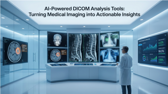
(DICOM) Digital Imaging and Communications in Medicine is the global standard used to store, transmit, and manage medical imaging data such as CT scans, MRIs, and X-rays. It ensures that imaging devices and software from different manufacturers can communicate and exchange data seamlessly.
By combining image recognition, automation, and intelligent reporting, modern AI DICOM analysis platforms are turning raw imaging data into structured, actionable information that supports faster and more confident clinical decisions.
AI-powered DICOM analysis tools use advanced AI analysis and image recognition technologies to assist radiologists in identifying patterns, measuring abnormalities, and generating faster reports.
Why DICOM Analysis Needs to Evolve
The DICOM standard has made it possible to store and share medical images across different systems and institutions. However, most DICOM viewers were originally designed for display, not for advanced analysis. As imaging technology improved, the volume of data contained in each study increased significantly.
A single CT scan today may contain hundreds of slices. MRI studies can include multiple sequences. Ultrasound exams often require repeated measurements and annotations. Reviewing all this data manually takes time, and even experienced radiologists can be slowed down by the sheer amount of information.
In busy radiology departments, this leads to longer reporting times, higher fatigue, and greater risk of inconsistency. The need for tools that can assist with analysis without replacing clinical judgment has become clear.
AI-enabled DICOM analysis tools address this gap by helping radiologists identify, measure, and organise imaging findings more efficiently.
How AI Improves Radiology Image Analysis
Modern AI diagnostic imaging software like RadpicsAI uses image recognition and machine learning models trained on large datasets to detect patterns within medical images. These tools do not make clinical decisions on their own, but they can highlight structures, measure abnormalities, and prepare information for the radiologist to review.
For example, AI can automatically identify nodules, fractures, or areas of abnormal density within a scan. It can calculate dimensions, volumes, or angles without requiring manual input. Instead of performing repetitive measurements, the radiologist can verify the results and focus on interpretation.
This approach significantly reduces the time required to complete a case while maintaining full clinical control.
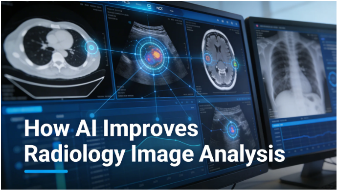
AI-powered analysis also helps standardise reporting. When measurements and annotations follow consistent rules, reports become easier to read and compare over time. This improves communication between radiologists, clinicians, and surgeons.
From Image Viewing to Actionable Insights
One of the biggest advantages of AI radiology analysis is the ability to convert visual data into structured information. Traditional workflows often separate image viewing from report creation, forcing radiologists to switch between tools.
With an integrated medical imaging AI platform like RadpicsAI, analysis happens within the same environment as reporting. Findings detected by AI can automatically appear in the report template. Measurements can be inserted directly into structured fields. Annotations remain linked to the original image.
This reduces duplication of work and ensures that the final report accurately reflects what is seen on the scan.
In high-volume settings, these small improvements add up quickly. Faster analysis leads to faster reporting, which improves turnaround time and allows radiology teams to handle more cases without increasing workload.
Consistency and Accuracy in High-Volume Radiology
As imaging demand grows, maintaining consistency becomes as important as speed. Manual measurements can vary between operators, especially when workloads are heavy. Even small differences can affect follow-up decisions or treatment planning.
Automated radiology AI tools help reduce this variation by applying the same measurement and detection rules every time. This does not replace the radiologist's judgment, but provides a reliable starting point.
Standardised analysis also makes it easier to compare studies over time. When previous measurements are stored in a structured format, follow-up scans can be reviewed more efficiently, improving long-term patient care.
Hospitals and diagnostic centres benefit from this consistency because reports become more predictable, easier to audit, and easier to share across departments.
Supporting Remote and Multi-Site Reporting
Modern healthcare workflows often involve multiple locations. Radiologists may report remotely, diagnostic centres may share cases with specialists, and hospitals may rely on external reporting teams during peak hours.
AI-enabled DICOM analysis platforms make collaborative and accessible medical image analysis possible by providing cloud-based access to imaging data and AI analysis tools without requiring complex local installations. Measurements, annotations, and reports remain synchronised across users, allowing radiology teams to collaborate efficiently without workflow delays
This flexibility is especially important for tele-radiology and large hospital networks, where efficiency depends on smooth communication between sites.
The Future of AI in Radiology Workflow
AI in radiology is still evolving, but the direction is clear. Analysis tools will continue to become more accurate, more integrated, and more supportive of clinical workflows. Future systems will not only detect abnormalities but may also assist with case prioritisation, quality checks, and decision support.
The goal is not to replace radiologists, but to give them tools that reduce repetitive work and allow more attention to be spent on complex interpretation.
As imaging volumes continue to grow, the ability to turn DICOM data into meaningful insights quickly will be essential for maintaining both speed and quality.
Conclusion
AI-powered DICOM analysis tools are changing the way medical imaging is interpreted. By combining image recognition, automated measurements, and integrated reporting, these systems help radiology teams work more efficiently while maintaining clinical accuracy.
In an environment where demand continues to rise, intelligent analysis platforms make it possible to convert large volumes of imaging data into actionable insights without increasing the burden on radiologists
Frequently Asked Questions
What is a DICOM analysis tool?
A DICOM analysis tool is software that helps radiologists interpret medical images stored in the DICOM format. These tools allow users to view, measure, annotate, and analyze imaging data from modalities such as CT, MRI, X-ray, and ultrasound. Modern AI-powered DICOM analysis tools can automatically detect abnormalities, calculate measurements, and assist in preparing structured radiology reports.
How does AI improve DICOM image analysis in radiology?
AI improves DICOM image analysis by using machine learning and image recognition algorithms to identify patterns within medical images. AI-powered systems can highlight potential abnormalities, perform automated measurements, and assist with image segmentation. This reduces manual work for radiologists and helps improve reporting speed, accuracy, and consistency.
Can AI-powered DICOM analysis tools replace radiologists?
No. AI-powered DICOM analysis tools are designed to support radiologists, not replace them. These tools assist with repetitive tasks such as measurements, detection, and annotation, allowing radiologists to focus on clinical interpretation and decision-making. Final diagnosis and reporting always remain under the radiologist's control.
How do DICOM analysis tools integrate with PACS systems?
Most modern DICOM analysis tools integrate directly with PACS (Picture Archiving and Communication Systems). This integration allows radiologists to analyze images, perform measurements, and generate reports within the same workflow without switching between multiple systems. PACS integration helps improve efficiency and reduces reporting time.
What are the benefits of AI-powered DICOM analysis for hospitals and diagnostic centers?
AI-powered DICOM analysis tools help healthcare organizations improve radiology workflows by reducing reporting time, increasing diagnostic consistency, and supporting collaboration between radiologists. These systems also help manage high imaging volumes, improve reporting turnaround times, and enhance the overall efficiency of medical imaging departments.
What is the difference between a DICOM viewer and a DICOM analysis tool?
A DICOM viewer allows clinicians to open and visualize DICOM images, such as CT, MRI, or X-ray scans. A DICOM analysis tool goes further by providing advanced capabilities like automated measurements, lesion detection, image segmentation, and AI-assisted analysis. While viewers focus on visualization, analysis tools help extract clinical insights from medical images.
How does AI help reduce radiology reporting time?
AI helps reduce radiology reporting time by automating repetitive tasks such as image measurements, lesion detection, and report structuring. Instead of manually calculating values or identifying regions of interest, radiologists can review AI-generated insights and focus on interpretation. This improves workflow efficiency and allows radiology teams to handle higher imaging volumes without increasing workload.
Why are AI-powered medical imaging platforms becoming important in modern healthcare?
AI-powered medical imaging platforms are becoming important because imaging volumes are increasing rapidly while the number of radiologists remains limited. These platforms combine PACS, DICOM analysis tools, and AI algorithms to streamline workflows, improve diagnostic consistency, and support faster clinical decisions. By turning complex imaging data into structured insights, AI platforms help healthcare systems deliver more efficient and scalable diagnostic services.